Search
- Page Path
- HOME > Search
Review Article
- Beyond the Number: Interpreting Prostate-Specific Antigen Elevation in the Context of Prostate Inflammation
- Byoungkyu Han, Ki-Hyuck Moon
- Urogenit Tract Infect 2025;20(3):132-143. Published online December 31, 2025
- DOI: https://doi.org/10.14777/uti.2550032016
-
 Abstract
 PDF
PDF Supplementary Material
 PubReader
 ePub
ePub - Prostate-specific antigen (PSA) is indispensable but not cancer specific; inflammation, benign prostatic hyperplasia, urinary retention, ejaculation, and instrumentation can all elevate PSA and complicate cancer risk assessment. This review synthesizes current evidence and guidelines to support clinicians in interpreting PSA elevations when inflammation is present or suspected. Acute febrile urinary tract infection and acute bacterial prostatitis may produce very high PSA values, sometimes exceeding 100 ng/mL, and normalization can be slow; therefore, PSA testing during active infection is discouraged. When PSA is only mildly to moderately elevated, standardized repeat testing is essential because a meaningful proportion of results normalize on retesting. A magnetic resonance imaging (MRI)-first pathway improves detection of clinically significant prostate cancer while reducing overdiagnosis and enables biopsy deferral after a negative MRI under structured monitoring. PSA density (PSAD) further refines triage alongside MRI, with practical working thresholds of roughly 0.10–0.20 ng/mL/cm3 calibrated to MRI quality and pretest risk. However, asymptomatic histologic prostatitis (National Institutes of Health category IV) is common and may raise PSA without reliably altering PSAD, which means that PSAD alone cannot confirm that an elevation is attributable solely to inflammation. Validated secondary biomarkers (e.g., Prostate Health Index, 4Kscore, IsoPSA [isoform PSA], Stockholm3, Proclarix, PCA3 [prostate cancer gene 3], SelectMDx [select molecular diagnostics], ExoDx [exosome diagnostics], MPS/MPS2 [MyProstateScore/MyProstateScore 2.0]) are best used selectively when MRI is negative or equivocal and clinical risk remains uncertain. A pragmatic sequence—confirm, image, and refine—helps minimize missed clinically significant cancer while reducing unnecessary antibiotics and biopsies when inflammation is the predominant driver of PSA elevation.
- 386 View
- 13 Download

Case Report
- Hemangioma Mistaken for Renal Cell Carcinoma in a Patient With End-Stage Renal Disease: A Case Report
- Hyung-Lae Lee, Dong-Gi Lee, Jeong Woo Lee, Jeonghyouk Choi
- Urogenit Tract Infect 2025;20(1):48-51. Published online April 30, 2025
- DOI: https://doi.org/10.14777/uti.2550008004

-
 Abstract
 PDF
PDF PubReader
 ePub
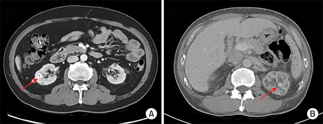
ePub - Hemangiomas are rare, benign vascular neoplasms that are more common in patients with end-stage renal disease. Here, we describe 2 cases of hemangioma misdiagnosed as renal cell carcinoma before renal transplantation. The key finding in our case was the misdiagnosis of hemangiomas as renal cell carcinoma based on computed tomography and magnetic resonance imaging in patients with end-stage renal disease. Because living transplantation was planned for our patients, we performed rapid surgical resection of the heterogeneously enhancing renal masses to avoid delays in transplantation. Our case highlights the importance of rapid surgical resection of enhanced renal masses to confirm diagnosis, thereby avoiding delays in patients scheduled for renal transplantation.
-
Citations
Citations to this article as recorded by- Editorial for UTI 2025 Vol. 20 No. 1 - Highlights of This Issue’s Papers and the UTI Editors’ Pick
Koo Han Yoo
Urogenital Tract Infection.2025; 20(1): 1. CrossRef
- Editorial for UTI 2025 Vol. 20 No. 1 - Highlights of This Issue’s Papers and the UTI Editors’ Pick
- 1,815 View
- 18 Download
- 1 Crossref

Review Article
- The Necessity of Human Papillomavirus Vaccination in Men: A Narrative Review
- Sooyoun Kim, Sangrak Bae
- Urogenit Tract Infect 2024;19(3):51-59. Published online December 31, 2024
- DOI: https://doi.org/10.14777/uti.2448030015

-
 Abstract
 PDF
PDF PubReader
 ePub
ePub - Anogenital wart caused by human papillomavirus (HPV) is the most common sexually transmitted infection. High-risk strains, such as types 16 and 18, cause penile cancer in men, cervical and vulvar cancers in women, and head and neck cancers and anal cancer in both sexes. Since these malignant tumors can be prevented through vaccination, the importance of vaccination is emphasized. However, because HPV is known to cause cervical cancer, vaccination is only being administered to women. Some countries vaccinate men as well, but in South Korea, only girls are included in the National Immunization Program. However, screening for HPV in men is not possible, and the virus causes various malignant tumors, with a sharp increase in head and neck cancers, as well as a surge in genital warts in the country. In addition, HPV worsens sperm quality. Moreover, the need for vaccines is increasing as the known methods for preventing HPV-related diseases in men are decreasing and the disease burden is increasing. As cost-effectiveness studies have shown that the cost-effectiveness of vaccination is lower for men than for women, it is unlikely that male vaccination will be included in national immunization programs. Many countries overseas, especially a very small number of OECD (Organization for Economic Cooperation and Development) countries including South Korea, are implementing mandatory vaccination for women. Vaccinating men and women, would be cost-effective and efficient in achieving herd immunity. In addition to herd immunity, the inclusion of male vaccination in the National Immunization Program is imperative given the rapidly increasing incidence of diseases in men.
-
Citations
Citations to this article as recorded by- Prevalence of sexually transmitted infections among persons aged 15–24 in Republic of Korea: A retrospective population-based descriptive study
Jinhee Seo, Minji Han, Sangrak Bae, Sooyoun Kim
Investigative and Clinical Urology.2026;[Epub] CrossRef - Human Papillomavirus Infection and Vaccine Uptake Among Males in the United Arab Emirates and the Wider Middle East and North Africa Region: A Narrative Review
Humaid AlKaabi, Raya Abu-Khalaf, Sandra Abu-Khalaf, Layan Abu-Khalaf, Yahia Khalil
Cureus.2025;[Epub] CrossRef
- Prevalence of sexually transmitted infections among persons aged 15–24 in Republic of Korea: A retrospective population-based descriptive study
- 10,826 View
- 107 Download
- 2 Crossref

Case Report
- Giant Fibroepithelial Polyp in the Renal Pelvis to the Upper Ureter
- Kyung Jin Chung
- Urogenit Tract Infect 2023;18(3):119-122. Published online December 31, 2023
- DOI: https://doi.org/10.14777/uti.2023.18.3.119

-
 Abstract
 PDF
PDF PubReader
 ePub
ePub - Benign ureteral tumors are rare owing to the predominance of malignancies in ureter tumors. Among them, fibroepithelial polyps are very rare, and giant fibroepithelial polyps are even rarer. This paper reports the author’s experience of giant fibroepithelial polyps detected incidentally. A 47-year-old woman was referred for severe right flank pain to the author’s center. A kidney computed tomogram revealed a long soft tissue lesion in the right kidney, upper polar calyx, pelvis, and whole proximal ureter with hydronephrosis. The radiologic reading was right renal pelvis urothelial cancer. Endoscopic removal was deemed impossible because the mass was filling the entire renal pelvis and upper ureter, so a nephroureterectomy was performed. After the nephroureterectomy, the pathology findings revealed a giant fibroepithelial polyp. This paper reports the treatment of giant fibroepithelial polyps of the ureter. Clinicians should consider the possibility of benign tumors and recommend a cautious approach to treatment if discovered.
- 2,570 View
- 17 Download

Original Article
- Predictor for Ureteral Stent Removal in Gynecological Cancer Patients with a Ureteral Obstruction
- Sung Hyun Ji, Young Kwon Hong, Young Dong Yu, Seung-Ryeol Lee
- Urogenit Tract Infect 2022;17(3):103-109. Published online December 31, 2022
- DOI: https://doi.org/10.14777/uti.2022.17.3.103
-
 Abstract
 PDF
PDF PubReader
 ePub
ePub - Purpose: This study investigated the possibility of ureteral stent removal in patients with gynecological cancer and ureteral obstruction.
Materials and Methods: From February 2010 to March 2022, the records of 121 patients with gynecological cancer and ureteral obstruction undergoing periodic ureteral stent replacement were reviewed retrospectively. Ureteral stent removal was attempted in patients who showed improvement in hydronephrosis on abdominal computed tomography (CT) after ureteral stent insertion and who lost symptoms. Success was defined as not having to reinsert the stent because there were no uncomfortable symptoms. The following were evaluated as predictors for ureteral stent removal: pre-removal serum creatinine, hypertension, diabetes, asymptomatic bacteriuria, type of gynecologic malignancy, prior radiotherapy, hydronephrosis laterality, obstruction level, hydronephrosis etiology, ureteral stent size, and ureteral stent duration. The hydronephrosis changes on abdominal CT were also evaluated.
Results: Among 121 patients, 74 with 81 obstructed ureters were evaluated. Of the 81 ureters, successful removal occurred in 30 (37.0%). There were significant differences between the success and failure cases regarding asymptomatic bacteriuria, prior radiotherapy, and hydronephrosis etiology. Logistic regression analysis showed that asymptomatic bacteriuria and hydronephrosis etiology were significant predictors of ureteral stent removal (p<0.05). There were also significant differences in the hydronephrosis changes depending on asymptomatic bacteriuria and hydronephrosis etiology (p<0.05).
Conclusions: In patients with gynecological cancer who have a ureteral stent in place due to ureteral obstruction, ureteral stent removal may be considered in patients who do not have asymptomatic bacteriuria or other etiologies, such as external ureteral pressure or ureteral cancer metastasis.
- 2,438 View
- 3 Download

Case Report
- Von Brunn’s Nest in an Incidental Bladder Mass Found during Holmium Laser Enucleation of the Prostate
- Seungsoo Lee
- Urogenit Tract Infect 2020;15(1):13-15. Published online April 30, 2020
- DOI: https://doi.org/10.14777/uti.2020.15.1.13
-
 Abstract
 PDF
PDF PubReader
 ePub
ePub - A 62-year-old male with benign prostatic hyperplasia underwent holmium laser enucleation of the prostate. During surgery, a small nodular mass was found incidentally in the trigone of the bladder. The lesion was removed completely by a transurethral resection with a bipolar device. A pathology examination of the lesion indicated von Brunn’s nests.
- 3,345 View
- 23 Download


 KAUTII

 First
First Prev
Prev


