



Articles
- Page Path
- HOME > Urogenit Tract Infect > Volume 21(2); 2026 > Article
- Case Report Acute Obstructive Pyelonephritis With Bilateral Hutch Diverticula in an Adult: A Case Report
-
Kyu Hyoung Cho1
 , Gilho Lee1, Youn Soo Jeon2
 -
Urogenital Tract Infection 2026;21(2):136-139.
DOI: https://doi.org/10.14777/uti.2652024.012
Published online: August 31, 2026
1Department of Urology, Soonchunhyang University Gumi Hospital, Gumi, Korea
2Department of Urology, Soonchunhyang University Cheonan Hospital, Cheonan, Korea
-
Corresponding author: Youn Soo Jeon Department of Urology, Soonchunhyang University Cheonan Hospital, 31 Suncheonhyang 6-gil, Dongnam-gu, Cheonan 31151, Korea Email: ysurol@schmc.ac.kr
• Received: April 12, 2026 • Revised: May 12, 2026 • Accepted: June 18, 2026
Copyright © Korean Association of Urogenital Tract Infection and Inflammation
This is an open access article distributed under the terms of the Creative Commons Attribution Non-Commercial License (http://creativecommons.org/licenses/by-nc/4.0) which permits unrestricted non-commercial use, distribution, and reproduction in any medium, provided the original work is properly cited.
- 411 Views
- 8 Download


Abstract
- Hutch diverticula are congenital bladder diverticula arising at the ureterovesical junction, typically associated with pediatric vesicoureteral reflux (VUR). Acute obstructive pyelonephritis occurring in an adult with bilateral Hutch diverticula is rare. The presence of a periureteral diverticulum may complicate ureteral stent placement and interpretation of postprocedural imaging findings. We report a 39-year-old man who was diagnosed with bilateral Hutch diverticula while being treated for acute obstructive pyelonephritis caused by a 4-mm proximal right ureteral stone. Following urgent right ureteral stenting and antimicrobial therapy, the patient's symptoms resolved, and he was discharged after 10 days of hospitalization. A subsequent voiding cystourethrogram confirmed bilateral Hutch diverticula but did not demonstrate definitive VUR. The imaging and cystoscopic findings clearly demonstrated the anatomical relationship between the right ureteral orifice and the adjacent right diverticulum. In addition, the distal coil of the double-J stent was lodged within the diverticulum, mimicking upward migration of the stent on kidney-ureter-bladder radiography. This case highlights a technical and radiologic pitfall of ureteral stenting in a patient with bilateral Hutch diverticula and emphasizes the importance of careful anatomical assessment during endourological intervention.
HIGHLIGHTS
The distal coil of a double-J stent may lodge within a Hutch diverticulum and mimic stent migration on radiography.
INTRODUCTION
Congenital bladder diverticula, specifically Hutch diverticula, are mucosal herniations through a defect in the muscular layer at the ureterovesical junction (UVJ) [1]. This condition is primarily diagnosed in children, often presenting as recurrent urinary tract infections (UTIs) associated with vesicoureteral reflux (VUR) [2]. However, Hutch diverticula in adults are uncommon and may remain clinically silent until complications arise [3]. Furthermore, acute obstructive pyelonephritis occurring in adults with bilateral Hutch diverticula is rare. In such patients, identification of the ureteral orifice during urgent ureteral stenting may be challenging because of the altered periureteral anatomy. We report a rare case of acute obstructive pyelonephritis caused by a proximal ureteral stone in an adult with bilateral Hutch diverticula. The case highlights a technical and radiologic pitfall in which the distal coil of a double-J (D-J) stent became lodged within a diverticulum, mimicking upward migration of the stent.
CASE PRESENTATION
A 39-year-old man presented with a 3-day history of right flank pain and fever (38.0°C). He had a history of acute pyelonephritis during childhood. Initial laboratory findings revealed leukocytosis (12,260/μL) and an elevated C-reactive protein level (4.49 mg/dL). Serum creatinine was elevated at 3.91 mg/dL. Serum glucose and hemoglobin A1c levels were 426 mg/dL and 12.9%, respectively. Urinalysis showed pyuria, and urine culture grew Escherichia coli. A noncontrast computed tomography (CT) scan demonstrated a 4-mm stone in the right upper ureter, causing acute obstructive pyelonephritis. Additionally, severe renal atrophy with parenchymal scarring of the left kidney was incidentally noted (Fig. 1A). Bilateral saccular outpouchings were observed at the UVJ, suggesting bilateral Hutch diverticula (Fig. 2A). The patient was treated with intravenous ceftriaxone, and a D-J stent was urgently inserted on the day of presentation for urinary drainage. During cystoscopy, identification of the right ureteral orifice was challenging because no visible ureteral urine jetting was observed, likely owing to the acute obstruction. After resolution of fever and stabilization of the infection, extracorporeal shock wave lithotripsy was performed on hospital day 7. After 10 days of inpatient medical treatment, all clinical manifestations of acute obstructive pyelonephritis had resolved, and the patient was discharged. One week after discharge, follow-up noncontrast CT confirmed complete stone clearance. Subsequent imaging and cystoscopy revealed that the distal coil of the stent was lodged within the right diverticulum rather than the bladder lumen (Fig. 2B and C). The D-J stent was removed on the same day. One week later, a voiding cystourethrogram demonstrated bilateral Hutch diverticula, although definitive VUR was not identified (Fig. 1B). The UTI resolved without recurrence, renal function remained stable, and the patient was scheduled for follow-up every 6 months.
DISCUSSION
Hutch diverticula result from deficient detrusor muscle support of the intravesical ureter, which predisposes patients to VUR by shortening the intravesical ureteral tunnel [1,2]. Although Hutch diverticula are primarily recognized in childhood, some cases remain undiagnosed until adulthood [3,4]. The likelihood of VUR in Hutch diverticula may be influenced by the location of the ureteral orifice and the size of the diverticulum. In the present case, cystoscopic examination confirmed that the ureteral orifices on both sides were located along the diverticular rims rather than within the diverticula. The right diverticulum was larger than the left (Fig. 2A). These anatomical findings suggest that the relationship between the ureteral orifice and diverticulum may vary between sides and may not fully explain the marked left renal atrophy in this patient. Nevertheless, transient or resolved reflux during childhood remains a possible contributing factor. Because a Hutch diverticulum may distort the configuration of the ureteral orifice or displace its location, identifying the ureteral orifice during urgent ureteral stenting can be challenging. In the present case, identification of the right ureteral orifice was further complicated by the absence of visible ureteral urine jetting, likely due to acute ureteral obstruction. Careful inspection of the diverticular rim was required to identify the ureteral orifice and achieve successful stent placement. A key clinical finding in this case was the clear delineation of the anatomical relationship between the right ureteral orifice and the adjacent diverticulum. In addition, the distal coil of the D-J stent was lodged within the diverticulum, mimicking upward migration of the stent on kidney-ureter-bladder radiography. The redundant space within the diverticulum can accommodate the distal coil and may mimic stent malposition on plain radiography; theoretically, this could also affect drainage or contribute to stent-related symptoms, although this was not observed in the present case [5]. In conclusion, we reported a case of acute obstructive pyelonephritis in an adult with bilateral Hutch diverticula. This case highlights a potential technical and radiologic pitfall during ureteral stenting.
-
Grant/Fund Support
This study received no specific grant from any funding agency in the public, commercial, or not-for-profit sectors.
-
Research Ethics
Written informed consent for publication was obtained from the patient. This case report was exempted from review by the institutional review board (IRB) of Soonchunhyang University Gumi Hospital (IRB No. 2026-06-01).
-
Conflict of Interest
The authors have nothing to disclose.
-
Author Contribution
Conceptualization: KHC, GL; Data curation: KHC; Methodology: KHC, GL; Project administration: YSJ; Visualization: KHC; Writing - original draft: KHC; Writing - review & editing: KHC, GL, YSJ.
NOTES
Fig. 1.
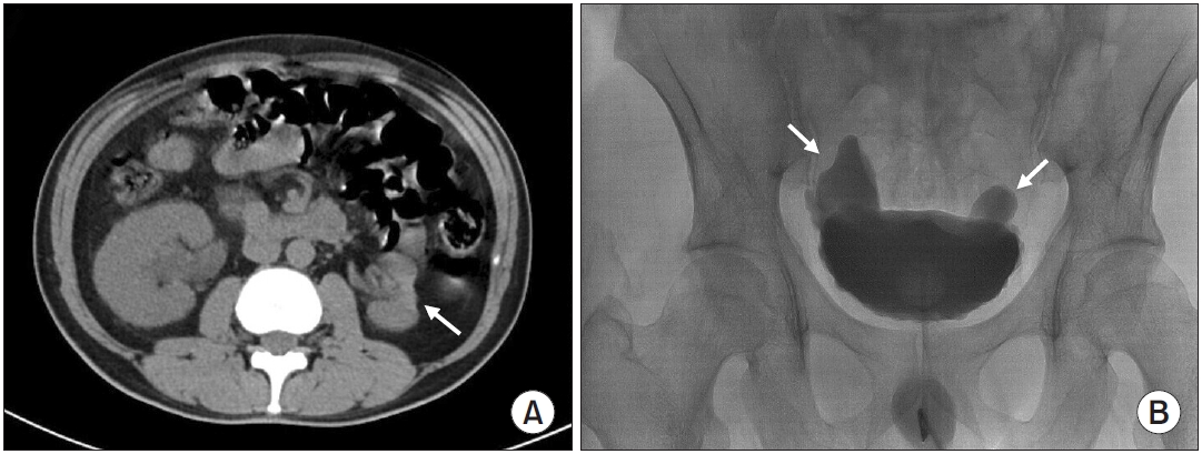
Imaging findings of Hutch diverticula and renal atrophy. (A) Axial noncontrast computed tomography image showing severe atrophy and parenchymal scarring of the left kidney (arrow). (B) Voiding cystourethrography demonstrating bilateral saccular outpouchings at the ureterovesical junctions (arrows), consistent with Hutch diverticula. No evidence of vesicoureteral reflux is observed.
Fig. 2.
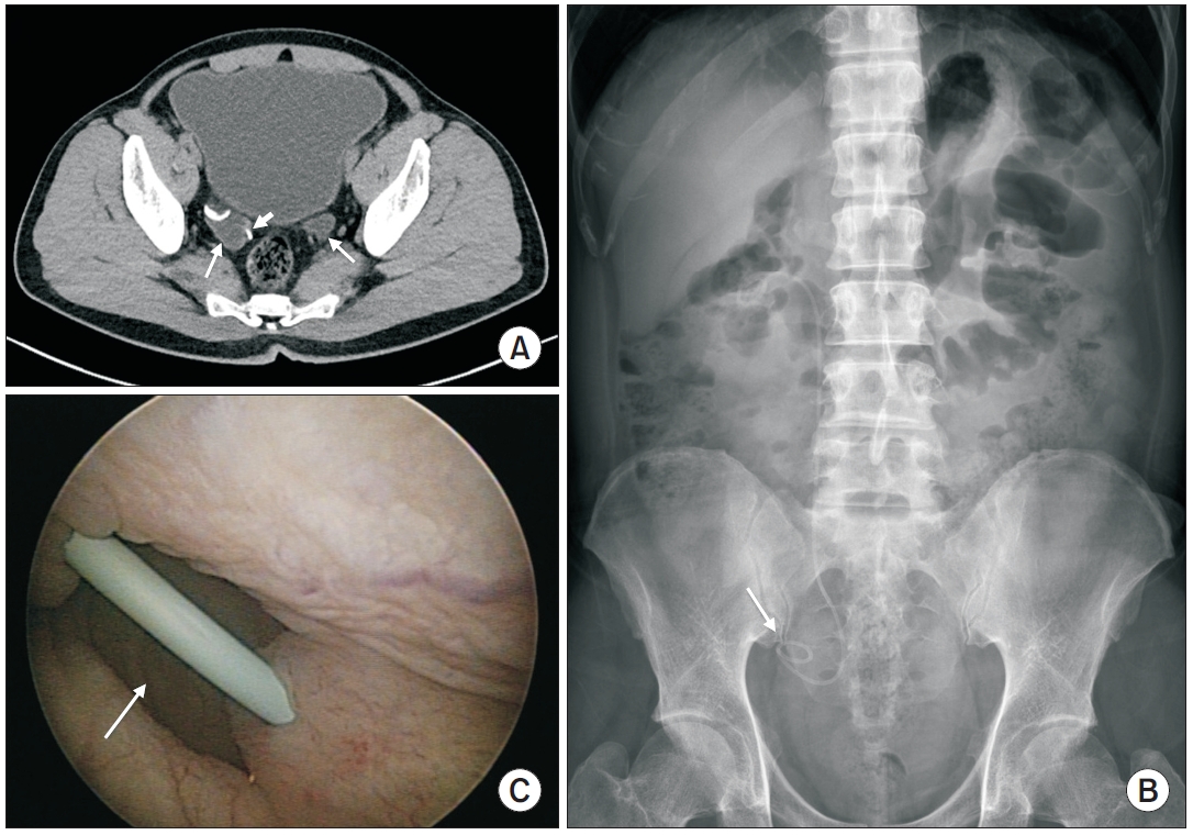
Radiologic and endoscopic findings of Hutch diverticula. (A) Axial noncontrast computed tomography (CT) image demonstrating bilateral Hutch diverticula (arrows) at the ureterovesical junctions. The distal right ureter courses adjacent to the right diverticulum, and cystoscopy confirmed that the right ureteral orifice was located along the diverticular rim. (B) Kidney-ureter-bladder radiograph demonstrating the distal coil of the double-J stent lodged within the right Hutch diverticulum (arrow), mimicking upward migration of the stent. (C) Cystoscopic image showing the opening of the right Hutch diverticulum (arrow). The diverticulum is sufficiently large to accommodate the distal segment of the double-J stent, corresponding to the CT and radiographic findings.
- 1. Hutch JA. Vesico-ureteral reflux in the paraplegic: cause and correction. J Urol 1952;68:457-69.ArticlePubMed
- 2. Garat JM, Angerri O, Caffaratti J, Moscatiello P, Villavicencio H. Primary congenital bladder diverticula in children. Urology 2007;70:984-8.ArticlePubMed
- 3. Fanning DM, Morrin M, Power R. An unusual presentation of bilateral Hutch diverticula. BMJ Case Rep 2014;2014:bcr2014204398.ArticlePubMedPMC
- 4. Yang CH, Lin YS, Ou YC, Weng WC, Huang LH, Lu CH, et al. Adult metaplastic Hutch diverticulum with robotic-assisted diverticulectomy and reconstruction: a case report. World J Clin Cases 2020;8:4895-901.ArticlePubMedPMC
- 5. Damiano R, Oliva A, Esposito C, De Sio M, Autorino R, D'Armiento M. Early and late complications of double pigtail ureteral stent. Urol Int 2002;69:136-40.ArticlePubMedPDF
REFERENCES
Figure & Data
REFERENCES
Citations
Citations to this article as recorded by 
 ePub Link
 Cite
Cite XML Download
Acute Obstructive Pyelonephritis With Bilateral Hutch Diverticula in an Adult: A Case Report
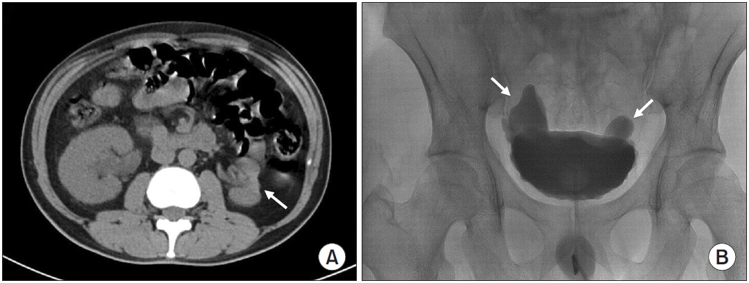
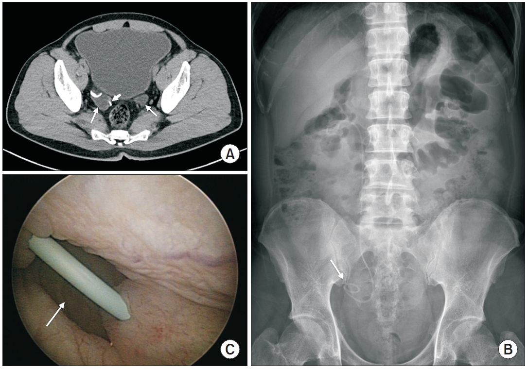
Fig. 1. Imaging findings of Hutch diverticula and renal atrophy. (A) Axial noncontrast computed tomography image showing severe atrophy and parenchymal scarring of the left kidney (arrow). (B) Voiding cystourethrography demonstrating bilateral saccular outpouchings at the ureterovesical junctions (arrows), consistent with Hutch diverticula. No evidence of vesicoureteral reflux is observed.
Fig. 2. Radiologic and endoscopic findings of Hutch diverticula. (A) Axial noncontrast computed tomography (CT) image demonstrating bilateral Hutch diverticula (arrows) at the ureterovesical junctions. The distal right ureter courses adjacent to the right diverticulum, and cystoscopy confirmed that the right ureteral orifice was located along the diverticular rim. (B) Kidney-ureter-bladder radiograph demonstrating the distal coil of the double-J stent lodged within the right Hutch diverticulum (arrow), mimicking upward migration of the stent. (C) Cystoscopic image showing the opening of the right Hutch diverticulum (arrow). The diverticulum is sufficiently large to accommodate the distal segment of the double-J stent, corresponding to the CT and radiographic findings.
Fig. 1.
Fig. 2.
Acute Obstructive Pyelonephritis With Bilateral Hutch Diverticula in an Adult: A Case Report


