Intraoperative Factors Associated With Postoperative Febrile Infectious Complications Following Retrograde Intrarenal Surgery: A Propensity Score-Matched Study
Article information

Abstract
Purpose
To investigate the intraoperative factors associated with postoperative febrile infectious complications following retrograde intrarenal surgery (RIRS).
Materials and Methods
A total of 352 patients who underwent RIRS for renal stones between March 2017 and December 2022 were included in the analysis. Preoperative factors included age, sex, body mass index, underlying diseases, preoperative urine analysis results, and stone characteristics on computed tomography scans. Patients were divided into 2 groups based on the presence (group B) or absence (group A) of postoperative febrile infectious complications. Intraoperative factors—such as the degree of hydronephrosis on retrograde pyelography (RGP), stone impaction on endoscopic findings, size of the ureteral access sheath (UAS), grade of ureteral wall injury, operation time, and stone composition—were compared retrospectively.
Results
Postoperative febrile infectious complications occurred in 44 patients (12.6%). Following 1:3 propensity score matching, preoperative factors were analyzed for 44 patients (group B) and 132 patients (group A). Group B had a significantly longer hospital stay than group A (p=0.007); however, no significant differences were observed in postoperative pain (p=0.862) or stone-free rate (p=0.197). Hydronephrosis (odds ratio [OR], 7.31; 95% confidence interval [CI], 2.65–20.19) and high-grade ureteral wall injury (OR, 4.15; 95% CI, 1.88–9.12) were independently associated with postoperative febrile infectious complications, whereas no significant associations were observed for stone impaction, UAS size, operation time, or infectious stone composition.
Conclusions
Hydronephrosis identified on RGP and high-grade ureteral wall injury during RIRS were associated with postoperative febrile infectious complications and may serve as potential markers for risk stratification.
HIGHLIGHTS
Hydronephrosis identified on retrograde pyelography and high-grade ureteral wall injury were independently associated with postoperative febrile infectious complications after retrograde intrarenal surgery. These intraoperative findings may provide real-time markers for postoperative infectious risk stratification. Incorporating surgeon-recognized intraoperative factors may improve early identification and surveillance of high-risk patients.
INTRODUCTION
Retrograde intrarenal surgery (RIRS) is a well-established minimally invasive treatment for upper urinary tract stones measuring less than 20 mm [1,2]. Advances in laser technology have enhanced the efficacy of RIRS and expanded its clinical application. Nevertheless, the overall complication rate following ureteroscopy ranges from 4% to 25%, with most being minor and self-limiting. Among these, postoperative infectious complications, including fever, systemic inflammatory response syndrome, upper and lower urinary tract infections (UTIs), hematuria, and postoperative pain, are relatively common [3-8].
Prophylactic antibiotics have shown efficacy in reducing the risk of infectious complications; however, despite identification and management of preoperative UTIs and careful perioperative planning in patients with multiple comorbidities, urosepsis remains a vital and potentially life-threatening complication, with an incidence of up to 5% [3-5]. Accordingly, the prevention and risk stratification of postoperative UTIs following RIRS have become important clinical challenges.
Therefore, this study aimed to investigate the association between intraoperative, surgeon-assessed real-time clinical factors during RIRS and the development of postoperative febrile infectious complications, and evaluate their impact on postoperative outcomes.
MATERIALS AND METHODS
1. Data Collection
With approval from the institutional review board (HDT 2025-11-003), data from patients who underwent RIRS with a ureteral access sheath (UAS) for renal stones between March 2017 and December 2022 were retrospectively analyzed. The requirement for informed consent was waived because of the retrospective nature of the study.
Clinical data were collected via a retrospective review of electronic medical records. Patients with a history of preoperative extracorporeal shock wave lithotripsy (ESWL), preoperative antibiotic treatment for a diagnosed UTI, previous ipsilateral ureteral surgery, or anatomical abnormalities, such as a horseshoe kidney or duplicated ureter, were excluded from the analysis. Perioperative antibiotic prophylaxis was administered in accordance with our institutional protocol, comprising intravenous antibiotics on the day of surgery and a single additional dose within 24 hours postoperatively. Postoperative febrile infectious complications were defined as fever ≥38.0°C persisting for more than 48 hours after surgery despite prophylactic intravenous antibiotic administration, or a positive postoperative urine culture. This composite definition was adapted from previous studies investigating infectious complications following ureteroscopic surgery [9-11]. As postoperative urine cultures were not obtained routinely but only in patients with clinically suspected UTI, postoperative febrile infectious complications were considered a composite clinical infectious outcome rather than culture-confirmed UTI alone. Patients were categorized into 2 groups according to the absence (group A) or presence (group B) of postoperative febrile infectious complications.
Preoperative factors included age, sex, body mass index (BMI), comorbidities (hypertension and diabetes mellitus), history of ESWL, Charlson Comorbidity Index (CCI), preoperative urine culture and urinalysis results, and stone-related parameters assessed using preoperative computed tomography, including stone size, multiplicity, and laterality. Intraoperative factors included the presence of hydronephrosis on retrograde pyelography (RGP), stone impaction on endoscopic evaluation, UAS size, grade of ureteral wall injury, operation time, and stone composition. Postoperative outcomes included length of hospital stay, postoperative pain requiring analgesics (defined as a visual analog scale score ≥ 4), and stone-free rate.
2. Surgical Technique
All procedures were performed by a single experienced surgeon with experience of over 300 surgeries for nephrolithiasis, using an 8.5F flexible ureterorenoscope. Under general anesthesia in the lithotomy position, a rigid ureteroscope was introduced to evaluate the lower urinary tract, followed by RGP. A safety guidewire (ZIPwire, Boston Scientific, USA) was inserted, and the ureter was endoscopically inspected. A 10F dual-lumen ureteral catheter was used to introduce a stiff guidewire (Amplatz Super Stiff, Boston Scientific), over which a UAS (UroPass, Olympus Europa SE & Co. KG, Germany) was advanced and positioned below the renal pelvis under fluoroscopic guidance. UAS size was determined according to intraoperative RGP findings and endoscopic assessment. At the end of the procedure, the UAS was removed under direct endoscopic visualization, and ureteral wall injury was graded [12]. A double-J stent was routinely placed and maintained for approximately one week postoperatively.
3. Statistical Analysis
Continuous variables were expressed as mean±standard deviation and compared using the independent-samples t-test. Categorical variables were presented as frequencies (%) and analyzed using the chi-square test or Fisher exact test, as appropriate. All statistical analyses were performed using R ver. 4.3.1 (R Foundation for Statistical Computing, Austria). Propensity score matching was conducted using 1:3 nearest-neighbor matching without replacement with the MatchIt package in R. Propensity scores were estimated using a logistic regression model based on preoperative variables. The propensity score model included age, sex, BMI, CCI≥3, and preoperative pyuria. Propensity score matching was performed without a caliper; no patients in group B were excluded during matching, only unmatched patients in group A were removed, and no missing data were present for variables included in the propensity score model. After matching, all standardized mean differences for the covariates were <0.1, and those for squared terms and 2-way interactions were <0.15, indicating satisfactory covariate balance. Preoperative variables and postoperative outcomes were compared between the matched groups. A 2-sided p-value <0.05 was considered statistically significant. To identify intraoperative factors associated with postoperative febrile infectious complications, logistic regression analysis was performed in the matched cohort. Variables significant on univariable analysis were entered into multivariable logistic regression with stepwise selection.
RESULTS
1. Preoperative Factors Associated With Postoperative Febrile Infectious Complications
Among the 352 patients who underwent RIRS, 2 with incomplete electronic medical records were excluded, leaving 350 patients for the final analysis. Postoperative febrile infectious complications occurred in 44 patients (12.5%). Baseline demographic, radiographic, and laboratory characteristics are summarized in (Table 1).
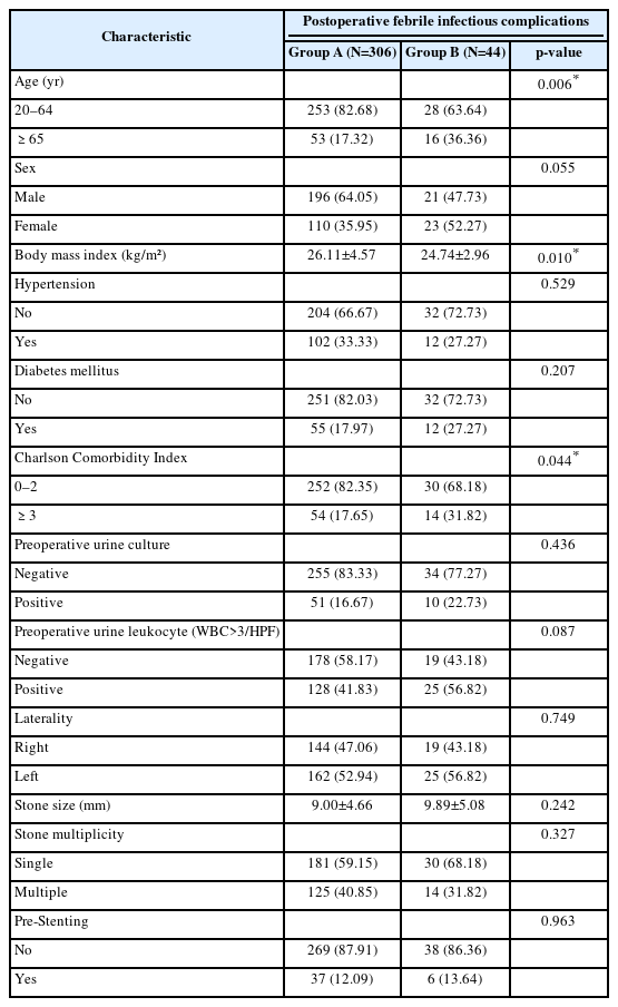
The preoperative characteristics of patients in groups A and B before propensity score matching
Patients aged ≥65 years old constituted a higher proportion in group B than group A (36.36% vs. 17.32%, p=0.006). BMI was significantly lower in group B (24.74± 2.96 kg/m² vs. 26.11±4.57 kg/m², p=0.010), and a CCI ≥ 3 was more frequent in group B (31.82% vs. 17.65%, p=0.044). (Fig. 1) illustrates the overall study design and flow chart of our research. A total of 12 preoperative factors were evaluated: 5 with p<0.1—age (p=0.006), sex (p=0.055), BMI (p=0.010), CCI ≥3 (p=0.044), and preoperative pyuria (p=0.087)—were selected for the 1:3 propensity score matching to adjust for baseline differences (Fig. 2). Following matching, 44 patients in group B and 132 patients in group A were included in the matched cohort (Table 2).
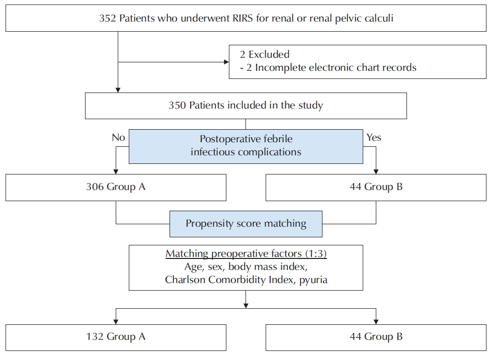
Flowchart of the study design. RIRS, retrograde intrarenal surgery; Group A, absence of postoperative febrile infectious complications; Group B, presence of postoperative febrile infectious complications.
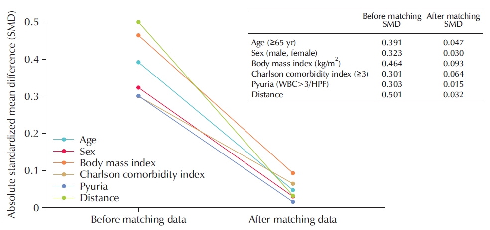
Covariate balance assessment before and after propensity score matching using standardized mean differences. WBC, white blood cell; HPF, high-power field.
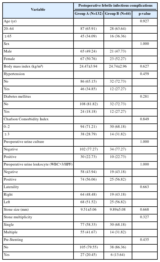
The preoperative characteristics of patients in groups A and B after propensity score matching
2. Intraoperative Factors Associated With Postoperative Febrile Infectious Complications
After propensity score matching, hydronephrosis identified on RGP was significantly associated with a higher incidence of postoperative febrile infectious complications than group A (29.55% vs. 6.06%, p<0.001). In addition, high-grade ureteral wall injury (grade≥2) was more frequent in group B (45.45% vs. 18.18%, p=0.001). No significant differences were observed between the groups in stone impaction, UAS size, operation time, or the presence of visually suspected infectious stones (Table 3). Multivariable logistic regression analysis demonstrated that hydronephrosis (odds ratio [OR], 7.31; 95% confidence interval [CI], 2.65–20.19; p<0.001) and high-grade ureteral wall injury (OR, 4.15; 95% CI, 1.88–9.12; p=0.001) were independently associated with postoperative febrile infectious complications (Table 4).
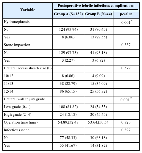
The intraoperative factors of patients in groups A and B after propensity score matching
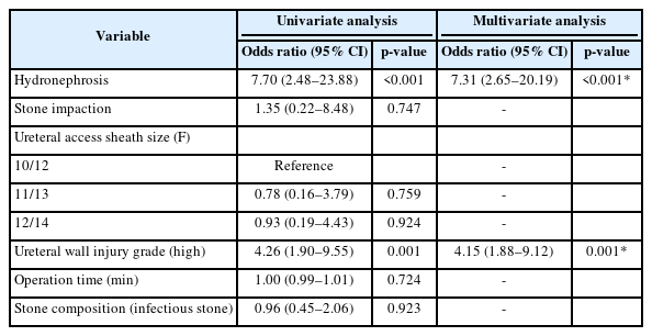
Intraoperative factors associated with postoperative febrile infectious complications in the matched cohort
3. Association Between Postoperative Outcomes and Postoperative Febrile Infectious Complications
Group B had a significantly longer hospital stay than group A (3.5±3.4 days vs. 1.9±1.8 days, p=0.007). In group B, 9 of 44 patients required escalation of antibiotic therapy for persistent fever, all of whom had positive postoperative urine cultures. Postoperative pain (p=0.862) and stone-free rate (p=0.197) did not differ significantly between the groups (Table 5).
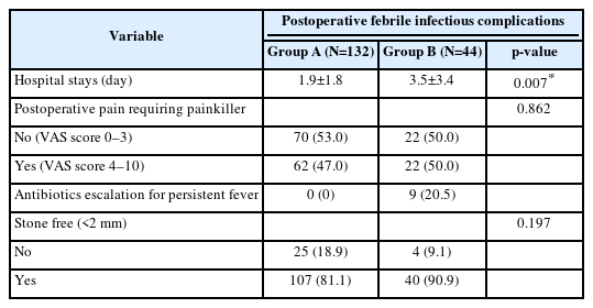
The postoperative outcomes of patients in groups A and B after propensity score matching
DISCUSSION
RIRS is recognized as a minimally invasive and effective treatment for renal stones; however, postoperative infectious complications remain a clinical concern. While ureteroscopy typically yields satisfactory outcomes with low morbidity, postoperative fever, UTI, and urosepsis continue to be relevant complications [3,4]. The incidence of post-ureteroscopy urosepsis ranges from 0.1% to 4.3%, while that of postoperative febrile infectious complications ranges from 0.2% to 15% of all reported cases [3-8]. To mitigate these infectious complications, UASs are widely used because they have been suggested to improve drainage and help maintain lower intrapelvic pressure during surgery; nevertheless, postoperative infectious complications are reported at a substantial rate despite adoption [4,13,14]. Previous studies have suggested several risk factors for infectious complications following RIRS, namely prolonged operation time, recent positive urine culture or UTI history, preoperative pyuria/nitrite, smaller-caliber UAS, struvite stones, high irrigation rate, and comorbidities [15]. Furthermore, one systematic review of sepsis following RIRS identified stone size, high irrigation pressure, prolonged stent dwelling time, positive urine culture, female sex, diabetes mellitus, and longer operative time as major risk factors [16]. However, most of these variables reflect preoperative patient characteristics or operative conditions, whereas intraoperative findings directly recognized by the surgeon have been less thoroughly evaluated.
Our findings have several important implications. First, postoperative febrile infectious complications following RIRS were associated with intraoperative findings that the surgeon could immediately recognize during the procedure. The postoperative febrile infectious complications group showed a higher frequency of hydronephrosis and a greater incidence of high-grade ureteral wall injury than the control group. In contrast, stone impaction, UAS diameter, operative time, and infectious stone composition showed no significant difference between the 2 groups. In addition, patients in the postoperative febrile infectious complications group had a longer hospital stay than controls, and antibiotics were escalated only in a subset of patients within the event group because of persistent fever (Table 5). These findings suggest that risk stratification for infectious complications following RIRS with UAS may be refined by incorporating intraoperative findings alongside conventional preoperative risk factors, thereby enabling more precise identification of patients at high risk of postoperative febrile infectious complications. Second, postoperative patients may present different symptoms and signs from those of patients with typical UTIs, and the use of prophylactic antibiotics may delay or obscure clinical or microbiological confirmation of infection. Therefore, patients with intraoperative risk findings require careful surveillance and appropriate antibiotic management in the immediate postoperative period [16]. In this regard, real-time intraoperative assessment may serve as an important tool for early monitoring and treatment of postoperative UTI and urosepsis.
Several mechanisms can explain the association between hydronephrosis and postoperative febrile infectious complications. Hydronephrosis suggests impaired drainage, urinary stasis, or functional obstruction of the upper urinary tract rather than representing a simple radiologic finding. Urinary obstruction and stasis are important pathophysiologic conditions predisposing the upper urinary tract to infection, as they may promote bacterial growth and persistence [17].
RIRS involves continuous irrigation and repeated instrumentation; therefore, dissemination of bacteria or inflammatory mediators can occur in a collecting system with impaired drainage. Therefore, the hydronephrosis observed herein may be interpreted not as a direct cause of infection, but rather a clinical marker of an upper urinary tract environment vulnerable to postoperative infection. Despite having previously been evaluated as a preoperative imaging factor, our findings suggest that hydronephrosis identified intraoperatively on RGP may provide clinically meaningful data for predicting postoperative febrile infectious complications [18].
The link between ureteral wall injury grade and postoperative febrile infectious complications can be understood in terms of access-related tissue injury and disruption of the urothelial barrier. UAS insertion and repeated ureteroscope manipulation can cause ureteral wall injuries, ranging from superficial mucosal damage to deeper injury involving the muscle layer. Previously proposed grading systems enable the systematic assessment of such injuries. Ureteral wall injury is associated with local inflammatory responses and ischemic changes [12,19]. Accordingly, the high-grade ureteral wall injury in our study suggests disruption of the ureteral tissue barrier, thereby creating a milieu more susceptible to postoperative infection. Such injury may contribute to postoperative fever through an acute inflammatory response and facilitate the clinical manifestation of pre-existing bacteria or infectious material via a disrupted urothelial barrier. A consensus document on ureteric injury likewise acknowledges ureteroscopy as a cause of iatrogenic ureteric injury. It highlights the importance of urine culture, consideration of antibiotics, and adequate drainage and stenting when ureteric injury is suspected or identified [20]. In this context, high-grade ureteral wall injury could serve as a marker of tissue vulnerability to postoperative infectious complications.
This study has several limitations. First, this was a retrospective single-center study performed by a single surgeon, which may limit generalizability and introduce selection bias. Second, although propensity score matching was used to reduce baseline differences between groups, the matched nature of the data was not explicitly accounted for in the regression analysis. Therefore, the observed associations should be interpreted with caution. In addition, residual confounding from unmeasured variables cannot be completely excluded. Third, we could not directly assess the effect of increased intrarenal pressure on the development of postoperative fever or UTI in individual patients. Although intrarenal pressure is an important intraoperative factor linked to infectious complications during RIRS [21-23], its real-time measurement remains limited in routine clinical practice. Accordingly, all procedures in our cohort were performed under standardized conditions, with the same irrigation settings and without a suction-access sheath. Future prospective studies incorporating intrarenal pressure or irrigation-related parameters, along with standardized outcome definitions, remain warranted.
Despite these limitations, this study is meaningful in that it evaluated surgeon-recognized intraoperative findings rather than relying solely on conventional preoperative predictors. Our findings suggest that real-time intraoperative observations during RIRS may provide additional clinical information for risk stratification of postoperative febrile infectious complications.
CONCLUSIONS
Hydronephrosis identified on RGP and high-grade ureteral wall injury during RIRS were associated with postoperative febrile UTI and may serve as potential markers for future risk stratification, underscoring the importance of meticulous intraoperative assessment. Prospective studies using standardized definitions of postoperative infectious complications are needed to validate these findings.
Notes
Grant/Fund Support
This study received no specific grant from any funding agency in the public, commercial, or not-for-profit sectors.
Research Ethics
This study was approved by the institutional review board (IRB) of Hallym University Dongtan Sacred Heart Hospital (approval number: HDT 2025-11-003). The requirement for informed consent was waived from the IRB because of the retrospective nature of the study.
Conflict of Interest
The authors have nothing to disclose.
Author Contribution
Research conception & design: JK Kim, Seong Ho Lee; Acquisition of data: Wongjong Yang, Sangyoug Park, Wonchul Lee; Data analysis and interpretation: JK Kim, Wongjong Yang, Chagil Choi, Hyung-Jee Kim; Draft of the manuscript with figures and tables: JK Kim, Wongjong Yang; All authors reviewed the manuscript.